Article Text

Abstract
Introduction Non-expansile lung (NEL) is a common cause of talc pleurodesis (TP) failure in malignant pleural effusion (MPE), but is often occult prior to drainage. Reliable detection of NEL would allow patients to be allocated between intrapleural catheter (IPC) and TP. High pleural elastance (PEL) has been associated with NEL in observational studies. Pre-EDIT is a randomised feasibility trial of elastance-directed IPC or TP (EDIT) management using a novel, purpose-built digital pleural manometer (Rocket Medical, UK).
Methods and analysis Consecutive patients with MPE without prior evidence of NEL or preference for IPC will be randomised 1:1 between EDIT management and standard care (an attempt at TP). The primary objective is to determine whether sufficient numbers of patients (defined as 30 within 12 months (or 15 over 6 months)) can be recruited and randomised to justify a subsequent phase III trial testing the efficacy of EDIT management. Secondary objectives include safety, technical feasibility and validation of study design elements, including the definition of PEL using 4D pleural MRI before and after fluid aspiration. EDIT involves PEL assessment during a large volume pleural fluid aspiration, followed by an attempt at TP or placement of an IPC within 24 hours. Patients will be allocated to IPC if the rolling average PEL sustained over at least 250 mL fluid aspirated (PEL250) is ≥ 14.5 cm H2O/L.
Ethics and dissemination Pre-EDIT was approved by the West of Scotland Regional Ethics Committee on 8 March 2017 (Ref: 17/WS/0042). Results will be presented at scientific meetings and published in peer-reviewed journals.
Trial registration number NCT03319186; Pre-results.
- pleural disease
This is an Open Access article distributed in accordance with the Creative Commons Attribution Non Commercial (CC BY-NC 4.0) license, which permits others to distribute, remix, adapt, build upon this work non-commercially, and license their derivative works on different terms, provided the original work is properly cited and the use is non-commercial. See: http://creativecommons.org/licenses/by-nc/4.0/
Statistics from Altmetric.com
Strengths and limitations of this study
A prospective assessment of the feasibility of a novel, pleural elastance (PEL)-directed treatment pathway (EDIT management) for malignant pleural effusion.
It utilises purpose-built digital pleural manometry equipment allowing continuous intrapleural pressure recording, and derivation of PEL via a user-friendly interface.
It includes pre-aspiration and post-aspiration 4D pleural MRI to validate assumptions regarding anatomical changes during fluid aspiration, the computation of PEL and the origin of symptoms, such as cough and chest pain.
It is a single-centre feasibility study; not powered to evaluate the efficacy of EDIT management but will inform the design a larger future trial using a patient-centred primary outcome measure.
Introduction
Malignant pleural effusion (MPE) is a common clinical condition and frequently leads to disabling breathlessness. Survival in patients with MPE is notably heterogeneous and is determined primarily by tumour type. However, most patients survive for less than a year and in some, this may be limited to a few months.1 Efficient palliation of MPE symptoms is therefore a major priority for patients and their families. This is currently achieved either by admission to hospital for intercostal drain placement and an attempt at talc pleurodesis (TP) or outpatient placement of an indwelling pleural catheter (IPC).2 When successful, TP results in durable symptom control after a 4–7 day hospital stay, and obviates the need for intermittent, domiciliary drainage and the risk of ongoing complications including pleural infection. However, non-expansile lung (NEL) frequently complicates an attempt at TP and cannot be reliably predicted using baseline radiology or other parameters. Since an IPC will effectively palliate most MPEs, regardless of NEL,2 some centres advocate their use in all patients with MPE. However, an IPC is not acceptable to some patients. Anecdotally, this is particularly the case if immunosuppressive cancer therapy is planned, or their lifestyle or environment makes IPC management inconvenient or overly restrictive (eg, patients who enjoy swimming or reside in a warm climate).
Current guidelines direct clinicians to offer a choice between TP or IPC based on patient preference, given the equivalent efficacy of these approaches in two randomised controlled trials.3 However, this apparent equivalence cannot be generalised to patients with NEL, since the incidence of NEL was extremely low in these important studies, occurring in only 3/54 (6%) and 2/72 (3%) patients in TIME-2 and AMPLE, respectively.4 5 In clinical practice, the incidence of NEL is almost certainly higher; in audit data recently reported by our unit, NEL was identified in 15/65 patients with MPE (23%) who underwent complete pleural fluid drainage.6 In this series, NEL was also associated with a twofold to fourfold increase in all-cause mortality, which may explain the under-representation of NEL in the TIME-2 and AMPLE studies.6
In our view, the ideal MPE treatment pathway would incorporate an initial functional assessment to determine the probability of underlying NEL, given the profound impact this has on the decision-making required by current guidelines (TP or IPC). Such a pathway, if accurate and efficient, would maximise patient choice in those with expansile lung (who could reasonably choose between TP and IPC) and minimise the rate of futile TP attempts in patients with NEL (avoiding the associated risks, cost and inconvenience). The current randomised feasibility pre-EDIT trial has been initiated to assess the feasibility and inform the design of a future randomised phase III trial testing the efficacy of elastance-directed IPC or TP (EDIT) management. Pre-EDIT also includes a Treatment Preferences Survey (TPS) to more adequately record patient views regarding decision-making in MPE management.
Rationale for the pre-EDIT trial
Detection of NEL
Using routinely available clinical information NEL cannot be reliably detected prior to complete pleural drainage. A thick visceral peel encasing the lung may be seen on cross-sectional imaging in gross cases but is frequently absent. Following diagnostic or therapeutic pleural fluid aspiration, an ex-vacuo (hydro-) pneumothorax may be visible.2 However, this is also an insensitive sign, since a NEL may re-expand enough to appear expansile after removal of currently recommended volumes of fluid (< 1.5 litres). Even after complete drainage of MPE, we have recently reported considerable interobserver variation in radiographic detection of NEL.6 Sonographic findings, including heavy septation or visceral pleural thickening,7 or abnormal M-mode and speckle-tracking end-points,8 have recently been reported as potential NEL biomarkers, but have yet to be validated in large cohorts. Development of a reliable predrainage biomarker for NEL is therefore urgently required.
Pleural elastance (PEL) is an intrinsic property of the pleural cavity and is defined following thoracentesis by the change in intrapleural pressure (∆IPP) divided by the change in pleural cavity volume, which is assumed to be equal to the volume of fluid removed (∆VOUT).9 In previous observational studies, elevated PEL has been strongly associated with the presence of NEL and the occurrence of TP failure.10 However, previous studies have not attempted to allocate patients to TP or IPC using PEL measurements. For this to be feasible, safe and reliable, various technical difficulties regarding IPP measurement need to addressed, in addition to pathway and trial design issues that are summarised below.
Technical considerations in IPP measurement
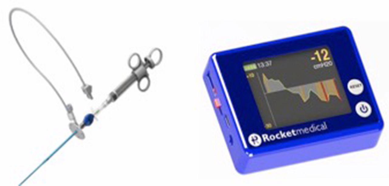
Previous pleural manometry equipment has been hampered by technical limitations, largely relating to poor damping of pressure variations related to normal respiration and/or the need for cumbersome improvised equipment. In conjunction with Rocket Medical (UK), we have developed a novel, single-use, CE-marked digital pleural manometry (DPM) catheter which allows continuous IPP measurement during thoracentesis (see figure 1). IPP is measured once per second and is mechanically damped via the narrow independent lumen linking the pleural cavity to the electronic transducer (ET). IPP is also temporally damped by displaying a mean IPP on a re-usable digital display unit, based on the preceding 5 s of data. The precision and accuracy of the electronic transducer within the catheter has been laboratory tested by Rocket Medical (UK) during product development and found to read within ±5% of a calibrated laboratory device at simulated pressures between +20 and −30 cmH2O. The manometer was EMC tested and passed BS EN 60601-1-2:2015 and BS EN 60601-1:2006+A1:2013.

Rocket Medical (UK) digital pleural manometer and re-usable display unit.
EDIT pathway design
Monophasic vs biphasic NEL and selection of an abnormal PEL threshold
Previous studies have reported characteristic patterns of IPP change in patients with NEL and expansile lung, during thoracentesis.9 11 12 Expansile lung is typically associated with a small linear reduction in IPP over the course of a large volume pleural aspiration. In contrast, NEL results in much larger fall in IPP, reflecting high PEL. This may occur early in the procedure where the aggregated compliance of the lung and parietal structures is severely limited resulting in a steep linear fall in IPP (monophasic NEL), typically in patients with gross visceral pleural thickening. In more subtle forms of NEL, IPP may initially behave similarly to expansile lung, as the pleural space partially conforms to the volume change and/or the lung expands as much as it can. However, this accommodation will eventually be overwhelmed as the lung reaches its maximum expansion, leading to a fall in IPP, but only after a considerable volume of fluid has been removed. This results in a biphasic PEL curve, which can only be detected if sufficient fluid is removed to reach the relevant inflection point. Importantly, TP success is likely to be reduced in patients with monophasic and biphasic NEL, and the EDIT pathway needs to be capable of detecting both.
In the only previous study that has linked PEL to TP success, Lan et al demonstrated that a PEL threshold of ≥ 19 cm H2O/L following aspiration of 500 mL of pleural fluid was associated with a sensitivity for NEL of 79% (95% CI 0.49 to 0.99).10 However, the sensitivity of this approach is likely to have been limited by selection of a PEL threshold significantly above the upper limit of normal, which was subsequently defined as 14.5 cm H2O/L,13 and the small aspiration volume used (500 mL), which is likely to have been inadequate to detect all cases of biphasic NEL. Adopting a lower PEL threshold and using a larger total aspiration volume may increase the sensitivity of NEL detection, but could potentially sacrifice specificity, particularly if IPP rises transiently due to coughing or unknown measurement artefacts.
Therefore, in the current pre-EDIT trial, we will use a novel definition of NEL, based on a rolling average of PEL recording over the preceding 250 mL fluid removed (PEL250), during a large volume pleural aspiration. The aspiration volume will only be limited by symptoms, a drop in IPP below previously reported safety thresholds or a target pleural effusion depth, based on repeated sonographic measurements (see Methods and analysis section for further detail). IPP pressures used to derive PEL will be consistently measured at end-expiration. NEL will be defined by a maximum PEL250 ≥ 14.5 cm H2O/L (the previously reported upper limit of the normal range for PEL)13 occurring at any point during large volume aspiration. This definition aims to detect NEL at the earliest possible opportunity, including in patients with biphasic NEL, while preserving specificity.
Delivery of elastance-directed management and safety considerations
By definition, the EDIT pathway requires an additional large volume thoracentesis prior to allocation to TP or IPC. If the allocated procedure cannot be delivered promptly, ideally on the same day, this offsets any pathway efficiency gained through detection of NEL. However, placement of any form of Seldinger drain may be technically challenging after removal of the majority of the effusion during PEL assessment. Therefore, within the EDIT protocol, thoracic ultrasound (TUS) images will be acquired regularly during aspiration, and the procedure terminated once a minimum safe depth of effusion has been reached (see Methods and analysis section for further details). The protocol also allows that, if required, a ‘Boutin-type’ needle can employed for pneumothorax induction to ensure safe placement of PEL-allocated IPC or TP. This is regularly practised at level II thoracoscopy centres, including our own, when no, or minimal, pleural fluid is present at local anaesthetic thoracoscopy.14 However, if the current study finds that this is frequently required to deliver EDIT management, this may impact on the feasibility of subsequent multicentre phase III trial and any subsequent clinical deployment.
Validation of the current definition of PEL using volumetric MRI
PEL is currently defined as ∆IPP (cm H2O)/∆VOUT (L), based on the assumption that ∆VOUT is equivalent to the underlying change in pleural cavity volume. However, this assumption may not be valid, due to a combination of air and local anaesthetic introduced during the procedure, variable compliance of the surrounding structures and transient parenchymal-pleural fistulation. Moreover, since ∆IPP describes the aggregated behaviour of a potentially biphasic process, PEL values may inadequately describe pleural physiology in this setting. This uncertainty will be explored in the secondary objectives using volumetric pleural MRI, which allows precise measurement of intrathoracic structures.15–17
If the current definition of PEL is validated using MRI, this may enhance the usability of the device used here. In the current protocol, ∆VOUT needs to be manually recorded in parallel with IPP and the data integrated post-procedure. In the future, real-time integration of validated ∆VOUT data with IPP data could facilitate real-time display of PEL (or PEL250), circumventing this time-consuming step. As exploratory objectives, volumetric MRI data will also be correlated with the development of symptoms during thoracentesis, the origin of which are poorly understood.18
Treatment preferences survey
All potentially eligible (pre-screened) patients will be asked to complete a TPS (see online supplementary appendix 1). These qualitative data will be of value in deciding whether to pursue future studies of EDIT management and optimising the design of these.
Supplementary file 1
Methods and analysis
Study design and setting
Pre-EDIT is a randomised feasibility trial. Thirty patients with symptomatic MPE will be recruited at a single centre: The Queen Elizabeth University Hospital, Glasgow, UK. Potentially eligible patients will also be identified and pre-screened at Glasgow Royal Infirmary. Patients will be randomised 1:1 to receive either pleural elastance-directed IPC or TP (‘EDIT management’), or standard care (placement of an intercostal drain and attempt at TP). A TPS will be offered to all potentially eligible (pre-screened) patients. The trial is sponsored by National Health Service (NHS) Greater Glasgow & Clyde and jointly funded by Rocket Medical (UK) and The West of Scotland Lung Cancer Research Group. The overall trial design is summarised in figure 2.

Pre-EDIT trial flow chart. EDIT, elastance-directed intrapleural catheter or talc pleurodesis; IPC, intrapleural catheter; IPP, intrapleural pressure; MPE, malignant pleural effusion; NEL, non-expansile lung; PEL, pleural elastance; TP, talc pleurodesis; TUS, thoracic ultrasound; ∆VOUT, volume of fluid removed.
Sample size
As a feasibility study, this trial is not subject to a formal sample size estimation. Recruitment of 30 patients is likely to allow a reasonable view of the barriers which might be met in delivering EDIT management, to explore possible solutions, to document the time required to deliver the procedure and to refine training. Previous estimates of NEL incidence indicate that each arm of the study is likely to contain only a small number of NEL cases which will clearly limit the conclusions that can be drawn regarding the chosen PEL250 threshold and MRI data. These have therefore been set as unpowered secondary and exploratory objectives. Instead, a particular focus will be the impact of the study design on recruitment rate, including the requirement for two pleural procedures and randomisation between two treatment arms. This will be assessed by careful review of (pre-) screening logs for consistent reasons for screen failure or unwillingness to participate.
Since pre-EDIT involves novel equipment and imaging protocols, a review of data completeness and quality will be undertaken after completion of TUS assessment, DPM and MRI scanning in the first five patients randomised to EDIT management. This initial experience may be used to refine trial-specific instructions (TSIs) for use in subsequent patients. If significant data are missing or data are of such poor quality as to be uninterpretable from these patients, recruitment may be extended by up to five patients to replace these.
Study objectives and outcome measures
Primary objective
The primary objective is to determine whether it is possible to recruit and randomise 30 patients over 12 months (or 15 patients in any 6-month period). The primary outcome measure will be recruitment rate.
Secondary and exploratory objectives
Secondary objectives and their associated outcome measures are summarised in table 1. Exploratory objectives are described in box 1.
Secondary objectives and associated end points in the pre-EDIT study
Exploratory objectives in the pre-EDIT study
To explore anatomical changes using MRI that may account for the development of symptoms such as chest pain or cough during large volume thoracentesis.
To explore the factors important to patients with symptomatic malignant pleural effusion when deciding upon first-line definitive pleural intervention.
To develop a novel predictive model for pleural effusion volume estimation applicable to a wide range of effusion volumes using uniplanar TUS measurement.
To evaluate the feasibility and potential utility of M-mode assessment of cardiac impulse lung displacement in patients undergoing EDIT management.
To investigate potential novel MRI biomarkers, such as visceral pleural thickness and parenchymal volume change and strain during respiratory cycle pre-DPM, which may predict NEL.
DPM, digital pleural manometer; EDIT, elastance-direct intrapleural catheter or talc pleurodesis; NEL, non-expansile lung; TUS, thoracic ultrasound.
Screening and eligibility assessment
Potentially eligible patients (defined as patients with symptomatic MPE) will be pre-screened by members of the parent clinical teams via cancer multidisciplinary team meetings, routine outpatient appointments and during inpatient reviews. Consecutive pre-screened patients meeting all inclusion criteria and without assessable exclusion criteria will be included in a pre-screening log, provided with a patient information sheet (PIS) at the earliest possible opportunity (see online supplementary appendix 2) and invited to a formal screening visit, during which all eligibility criteria will be assessed (see below). These patients will be given at least 24 hours between provision of the PIS and consent to consider participation.
Inclusion criteria
Clinically confident diagnosis of MPE, defined as any of the following:
Pleural effusion with histocytologically proven pleural malignancy OR
Pleural effusion in the context of histocytologically proven malignancy elsewhere, without a clear alternative cause for fluid OR
Pleural effusion with typical features of malignancy with pleural involvement on cross-sectional imaging
Degree of breathlessness for which therapeutic pleural intervention would be offered.
Age > 18 years.
Expected survival > 3 months.
Written informed consent.
Exclusion criteria
Women who are pregnant or lactating.
Clinical suspicion of NEL for which TP would not be offered.
Patient preference for first-line IPC insertion.
Previous ipsilateral failed TP.
Estimated pleural fluid volume ≤ 1 L (defined by TUS).
Any contraindication to ICD or IPC insertion, including:
Irreversible coagulopathy.
Inaccessible pleural collection, including lack of suitable IPC tunnel site.
Any contraindication to MRI scanning, including:
Claustrophobia.
Cardiac pacemaker.
Ferrous metal implants or retained ferrous metal foreign body.
Previously documented reaction to Gadolinium-containing intravenous contrast agent.
Significant renal impairment (estimated glomerular filtration rate < 30 mL/min).
Informed consent
Consent will be a two-step process. Pre-screened patients, potentially agreeable to trial involvement will be invited to a formal screening visit, during which a member of the research team will discuss the trial and seek written consent to formal screening (see online supplementary appendix 2). This will involve formal assessment of all eligibility criteria, including a bedside TUS for measurement of pleural effusion volume (VTUS). Patients attending a screening visit will be added to a screening log. Patients meeting all eligibility criteria will be asked to give written consent to trial enrolment and randomisation.
Treatment preferences survey
All pre-screened patients will be eligible and asked to participate in the TPS, irrespective of whether they meet all eligibility criteria and/or wish to participate in the main trial. Pre-screened patients will be provided with a separate TPS PIS (see online supplementary appendix 3) and will be given sufficient time, based on their own judgement, to consider participation before signing a separate consent form for involvement in the TPS (see online supplementary appendix 3).
Trial interventions
The trial interventions are summarised in figure 2 and described in detail below. Further information, in the form of TSIs for all interventions are available in online supplementary appendices 4–9.
Baseline assessments
Following consent, elective hospital admission and baseline assessment will be completed by a member of the trial team. The following data will be recorded prior to randomisation:
Patient demographics and physical characteristics.
Mode of presentation, current diagnosis and smoking history.
Eastern Cooperative Oncology Group performance status.
Medical history and current medication.
Previous pleural interventions.
Symptoms, including pain and breathlessness according to 100 mm visual assessment (VA) scores.
Results of routine haematological and biochemical tests (within 10 days).
Baseline TUS findings.
Randomisation
Immediately after baseline assessment, patients will be randomised 1:1 and allocated using random permuted blocks to either EDIT management or standard care. A validated online system will be used (www.sealedenvelope.com). The availability of potential minimisation factors for the subsequent phase III EDIT trial will be recorded but not used in randomisation, including the LENT prognostic score.1 The allocated management strategy will commence within 72 hours of randomisation.
Standard care
Following procedure-specific written consent, a 12Fr intercostal drain will be placed to facilitate passive fluid drainage at a rate not exceeding 1000 mL/hour. Chest radiographs (CXRs) will be performed post-ICD insertion and repeated every 18–24 hours following insertion. Four grams of sterile talc will be administered as a slurry, if there is no evidence of NEL or significant residual pleural fluid, as per existing guidelines.2 The intercostal drain will be removed once fluid output falls below 250 mL in the preceding 24-hour period, in the presence of a patent drain.
Where NEL or residual fluid is identified, drain patency will be assessed by flushing. Thoracic suction may be applied at the discretion of the primary physician. An additional CXR will be repeated after a further 18–24 hour period. Talc slurry will be administered once at least 50% re-apposition of the visceral and parietal pleural is achieved, based on visual CXR estimation, as per existing guidelines.2 If this is not achieved within 48 hours of drain placement, further management will be at the discretion of the primary physician. Further details are given in online supplementary appendix 4.
EDIT management
MRI scanning prior to large volume pleural fluid aspiration and PEL assessment
Patients will be scanned using a 3.0T Siemens Prisma MRI scanner. The affected thoracic cavity will be localised and an isotropic T1-weighted volume acquired using volumetric interpolated breath-hold examination (VIBE) sequences. A stack of axial slices covering the entire lung and surrounding pleura will be acquired as a set of short breath-holds. Time-resolved 3D MR imaging of the complete thorax will then be obtained during tidal free-breathing and maximal inspiratory/expiratory efforts. A modified time-resolved angiography with interleaved stochastic trajectories (TWIST) sequence will be used for this purpose. Following this, Gd-DTPA contrast (Gadovist) will be administered as a 15–40 mL bolus (0.05 mmol/kg). VIBE sequences will be reacquired at copied slice positions to provide comparative postcontrast images at multiple time points.19 Further scanning details are given in online supplementary appendix 5.
Thoracic ultrasound
Following MRI scanning, a TUS scan will be performed. This will allow the operator to identify a safe site for insertion of the digital pleural manometry catheter, where possible, in the posterior axillary line in the second rib space above the costophrenic angle. Deviation from this site will be recorded. For assessment of the secondary objectives, measurement of the lateral, posterior and median subpulmonic effusion heights in centimetres (LH, PH and SH) will be performed with the patient sitting upright at 90°. The total pleural effusion volume (in millilitres) will be estimated using the Goecke formula (VTUS = (LH +SH) x 70).20 For examination of the exploratory objectives (see box 1), M-mode measurements of the atelectatic lower lobe will also be acquired, during a breath hold at end-expiration.
Large volume pleural fluid aspiration and computation of PEL (PEL250)
After infiltration of the insertion site with local anaesthetic, the digital pleural aspiration catheter will be inserted at the site marked using TUS. Pleural fluid will be removed in 50 mL aliquots until any of the following occur:
The patient develops chest discomfort or excessive coughing.
An intrapleural pressure of ≤ −20 cmH2O is reached.
The target maximum aspiration volume is reached (horizontal costal to visceral pleural distance ≤ 30 mm).
Sequential end expiratory IPP measurements will be recorded after each 50 mL aliquot. Additionally, the highest and lowest IPP values recorded during maximal respiratory manoeuvres at 200, 500 and 1000 mL will be recorded. Postprocedure, PEL250 values will be calculated summarising PEL over the preceding 250 mL of fluid removed. The first PEL250 value will therefore be computed based on the IPP change between 0 and 250 mL divided by 0.25. Equivalent PEL250 values will then be computed after each subsequent 50 mL fluid removed. Thus, after 300 mL has been removed, the next PEL250 value will be computed using the IPP change between 50 and 300 mL, again divided by 0.25. The highest recorded PEL250 in each case (MaxPEL250) and total pleural fluid volume removed will be recorded. Further details are given in online supplementary appendix 6.
MRI scanning after large volume pleural fluid aspiration and PEL assessment
Following DPM, a further thoracic MRI scan will be performed. Identical T1-weighted VIBE and TWIST sequences will be acquired; however, further intravenous contrast will not be administered.
Allocation and delivery of PEL-directed management
Participants will be allocated to IPC or an ICD and attempt at TP based on their highest recorded PEL250 in each case (MaxPEL250), as follows:
MaxPEL250 ≥ 14.5 cm H2O/L: allocated to IPC
MaxPEL250 < 14.5 cm H2O/L: allocated to ICD and an attempt at TP
The allocated procedure must be completed within 24 hours of large volume pleural fluid aspiration and computation of PEL. Both will be performed using standard Seldinger techniques based on repeat TUS assessment, unless there is insufficient residual fluid left for this purpose (in the judgement of the operator). In this situation, a ‘Boutin-type’ needle may be used to obtain blunt access to the pleural cavity and create an iatrogenic (hydro-) pneumothorax for the purpose of safe drain insertion. The remainder of the ICD/IPC insertion will then proceed in the usual fashion. A CXR will be performed postprocedure to assess the drain position. Further detail is provided in online supplementary appendices 7–9.
Management following PEL-directed IPC or intercostal drain insertion
Patients allocated to IPC placement will be managed using standard local policies, as detailed in the TSI (see online supplementary appendix 9). For patients allocated to intercostal drain insertion and TP, postinsertion management will be identical to patients receiving standard care.
Data collection and study visits
VA scores
VA scores for chest pain and breathlessness will be documented at baseline and then daily for 7 days following ICD or IPC placement. Following this, VA scores will be performed weekly until the 28-day clinical follow-up appointment.
Study follow-up visits
Routine clinical appointments are planned for all patients at approximately 7, 28, 60 and 90 days after discharge. A single-study-specific follow-up visit at 90 days (±10) will coincide with routine follow-up where possible. At this visit, a CXR will be acquired and details of hospital admissions, repeat pleural interventions, clinic visits and survival status shall be recorded based on clinical history, augmented by electronic records systems. Where patients are unfit or unable to attend clinic, or have died, these data shall be recorded using electronic systems.
Further pleural intervention
In the event of hospital readmission, further pleural procedures will not be restricted by study participation. Where a gradual return of breathlessness is encountered during follow-up, the research team will assess the likely cause. IPC complications such as infection or blockage will be managed at the discretion of the primary physician. If recurrent MPE is identified following TP and occupies greater than an estimated one-third of the hemithorax on CXR, IPC insertion or therapeutic pleural aspiration will be offered if clinically appropriate. Intervention for smaller volume MPE recurrence will be offered where treatment consensus is achieved with a second respiratory physician blinded to trial group allocation. In the event of disagreement, a third (blinded) casting opinion will be sought.
Data management
Study data will be recorded on paper case report forms and securely transferred to the Queen Elizabeth University Hospital Clinical Research Facility and entered into a trial database. All CXR, CT and MRI images relating to trial participation will be securely stored on NHS systems in line with routine clinical practice. Representative TUS images and M-mode cine clips will be stored on an encrypted trial hard drive.
Statistical analysis plan
The primary objective of trial recruitment will be expressed as a mean monthly rate over the complete trial period and over each 6-month period during which the trial is open. The time taken to perform digital pleural manometry, its failure rate, the incidence of adverse events (AEs) and serious adverse events (SAEs) associated with EDIT management, the number of patients requiring a Boutin needle for ICD/IPC insertion and aspiration volume required to detect abnormal PEL will be reported by simple descriptive statistics or proportions where appropriate. Differences in mean/median values will be assessed using Students t-test or Wilcoxon rank test. Pearson χ2 test and exact 95% CIs will be used to compare proportions. A standard significance level of 0.05 will be used. The Bland-Altman method will be used to assess agreement between ΔVOUT and ΔVMRI measurements and between preaspiration VTUS and VMRI measurements.
End of trial
Trial recruitment commenced on 28 August 2017 and will terminate on 28 August 2018 or after 30 patients have been recruited, whichever is soonest. Trial participation will cease once the final patient has completed their 90-day follow-up.
Changes to the study protocol since trial opening
The protocol described accurately reflects V.2.5 of the protocol, dated 15 December 2017. The protocol history is summarised below:
V.2.0, dated 4 March 2017
At request of REC, recruitment process modified such that follow-up contact to invite for screening visit to be made by clinical nurse specialist rather than research team.
V.2.1, dated 5 April 2017
References to ‘Boutin needle’ changed to ‘Boutin-type needle’.
Randomisation process changed from minimisation using LENT score to simple random 1:1 allocation with recording of availability of LENT score to assess feasibility of minimisation by LENT in future EDIT trial.
V.2.2, dated 11 May 2017
Introduction of 5-patient ‘run-in’ period to allow refinement of TUS, DPM and MRI protocols and assessment of data completeness.
Removal of statement committing to guidewire placement down Boutin-type needle (where used).
Removal of statement committing to pneumothorax induction in endoscopy suite.
V.2.3, dated 4 August 2017
Extension of time allowed between randomisation and delivery of intervention from immediate to up to 72 hours.
V.2.4, dated 9 October 2017
Remove preprocedure TUS-estimated target volume as stop criterion for DPM and replace this with target horizontal costal-visceral pleural distance on TUS assessment at intervals during the aspiration procedure.
Removal of 2-week follow-up appointment.
Follow-up interval to start from date of discharge rather than date of procedure.
V.2.5, dated 15 December 2017
Recording of pre-screened patients.
Introduction of TPS.
Ethics and dissemination
Safety reporting
Details of any AEs or SAEs will be collected during routine and trial follow-up visits. All AEs and SAEs will be recorded in the patient’s medical records and reported to the trial sponsor.
Dissemination
Pre-EDIT, and the ultimate intention to definitively evaluate EDIT management if found to feasible, will be publicised at regional and national meetings. The results of pre-EDIT trial will be presented at scientific meetings and published in peer-reviewed journals.
Trial management
A Trial Management Group consisting of the chief investigator, clinical research fellow, lead trial research nurse, statistician and administrative assistant will oversee the running of the trial and meet on a monthly basis.
References
Footnotes
Contributors GAM: contribution to the conception and design of the work, data acquisition, analysis and interpretation of data for the work; drafting the work. GAM, ST, ACK, JEF, PM, AC, KGB: final approval of the version to be published; agrees to be accountable for all aspects of the work in ensuring that questions related to the accuracy or integrity of any part of the work are appropriately investigated and resolved. ST: contribution to the conception and design of the work and interpretation of data for the work. ST, ACK, JEF, PM, AC, KGB: revising the work critically for important intellectual content; final approval of the version to be published. ACK: contribution to the design of the work and data acquisition for the work. JEF: contribution to the design of the work, data acquisition, analysis, and interpretation of data for the work. PM: contribution to the design of the work, analysis and interpretation of data for the work. AC: contribution to the design of the work. KGB: principal contribution to the conception and design of the work; data acquisition, analysis and interpretation of data for the work
Funding This work was supported by Rocket Medical (UK) and the West of Scotland Lung Cancer Research Group (Award September 2015). KGB is part-funded by NHS Research Scotland.
Competing interests Rocket Medical (UK) have part-funded this work and will supply the digital pleural manometry equipment to be used in the study.
Patient consent Not required.
Ethics approval Pre-EDIT was approved by the West of Scotland Regional Ethics Committee on 8 March 2017 (Ref: 17/WS/0042).
Provenance and peer review Not commissioned; externally peer reviewed.