Article Text

Abstract
The BTS clinical statement for the diagnosis and management of ocular tuberculosis (TB) draws on the expertise of both TB and and ophthalmic specialists to outline the current understanding of disease pathogenesis, diagnosis and management in adults. Published literature lacks high-quality evidence to inform clinical practice and there is also a paucity of data from animal models to elucidate mechanisms of disease. However, in order to improve and standardise patient care, this statement provides consensus points with the currently available data and agreed best practice.
- tuberculosis
This is an open access article distributed in accordance with the Creative Commons Attribution Non Commercial (CC BY-NC 4.0) license, which permits others to distribute, remix, adapt, build upon this work non-commercially, and license their derivative works on different terms, provided the original work is properly cited, appropriate credit is given, any changes made indicated, and the use is non-commercial. See: http://creativecommons.org/licenses/by-nc/4.0/.
Statistics from Altmetric.com
Introduction
Tuberculosis (TB) is the clinical disease caused by infection with the intracellular bacillus Mycobacterium tuberculosis (Mtb). Ocular TB (OTB) represents a form of extrapulmonary TB, which can lead to visual loss from the irreversible destruction of intraocular tissues. While mycobacteria may be found in almost any tissue in or around the eye, the majority of OTB is intraocular and causes inflammation of the uvea (uveitis). This Clinical Statement is aimed at both the Respiratory/Infectious Disease physician looking after TB and Ophthalmic specialists to provide practice points for routine clinical practice in the management of OTB.
TB in general exists as a spectrum of infection and disease states, where a complex interaction between the host immune response, and the bacillus, determines the outcome.1 This can result in clearance of the bacillus, latent infection, subclinical disease and finally active disease, where the patient suffers symptoms in the affected organ, and the bacillus replicates freely (see table 1). OTB shares many of the characteristics of TB elsewhere in the body, and when Mtb causes direct infection of the eye, the predominant structures involved are the uvea and retina.2 Within this, there are a wide variety of ocular phenotypes reported, and the Collaborative OTB Study Nomenclature Working Group has attempted to provide an international consensus nomenclature to address this.3
Terminology
Typical phenotypes include:
Peripheral occlusive retinal vasculitis (Eales’ disease).
Choroidal granulomas.
Serpiginous or serpiginous-like chorioretinopathy.
‘Atypical’ phenotypes include a wide variety of uveitides from non-granulomatous anterior uveitis to intermediate uveitis and non-occlusive retinal vasculitis.
Importantly, however, and in contrast to the lungs, TB in the eye may present as a hypersensitivity state secondary to active disease elsewhere in the body, perhaps mediated by Mtb-specific lymphocytes cross-reacting to antigens in the eye.4–8 This, alongside the relative rarity of the disease in many healthcare settings (the worldwide prevalence of isolated OTB varies significantly from 0.2% to 10.5% of patients with uveitis depending on whether TB is endemic),9 10 and the relative difficulty of sampling the ocular structures to confirm microbiological evidence of TB disease, makes confident diagnosis of OTB particularly challenging.
Treatment of OTB may also be difficult given the concern around using oculotoxic drugs such as ethambutol, which form a mainstay of conventional TB therapy. Length of treatment may also be contentious given the possible involvement of the central nervous system (CNS), treatment of which is longer than that of disease elsewhere in the body.
Finally, a number of ophthalmic inflammatory conditions may be treated with immunosuppression, including anti-tumour necrosis factor alpha (anti-TNF) agents, which increase the risk of reactivation of TB.11 Some of these conditions may even resemble TB (eg, sarcoidosis), making decisions around treatment and immunosuppression particularly difficult.
Given this, both physicians and ophthalmologists diagnosing and treating OTB are left with a series of dilemmas. Is the eye problem they are faced with active TB, or an alternative inflammatory disease that requires immune suppression? If it is active TB, does it involve direct invasion and infection of the eye, or is it a hypersensitivity phenomenon? Or are they faced with a combination of latent TB infection (LTBI) (with the potential for reactivation) and a second and separate ocular disease?
This Clinical Statement for TB and Ophthalmic specialists therefore brings together uveitis and TB experts to outline the current understanding of disease pathogenesis, diagnosis and management in adults. Published literature lacks high quality evidence to inform clinical practice and there is also a paucity of data from animal models to elucidate mechanisms of disease. However, in order to improve and standardise patient care, this Statement will provide consensus points with the currently available data and agreed best practice.11–15
Given these difficulties, it is essential that patients with suspected OTB are managed jointly between specialists in TB and ophthalmology.
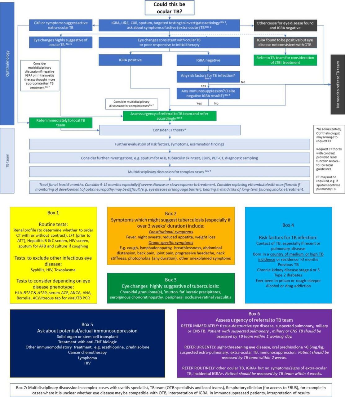
A management pathway is provided at figure 1.

Management pathway. AFB, acid–fast bacilli; ANA, antinucleaer antibody; ANCA, antineutrophil cytoplasmic antibody; ATT, antituberculous therapy; CNS, central nervous system; CXR, chest X-ray; EBUS, endobronchial ultrasound; IGRAs, interferon-gamma release-assays; LTBI, latent TB infection; OTB, ocular TB; PET, positron emission tomography; RCT, randomised controlled trial; TB, tuberculosis; TNF, tumour necrosis factor; U&E, urea and electrolytes.
Clinical practice point
All patients suspected of OTB should be managed jointly by ophthalmic specialists and TB centres.
Summary of clinical practice points
General
All patients suspected of OTB should be managed jointly by ophthalmic specialists and TB centres.
Epidemiology of OTB in adults and the effect of immunosuppression
Consider OTB as a cause of ocular disease particularly when individuals have risk factors for TB.
In immunosuppressed individuals, TB may be more disseminated and include the eye.
OTB may be present despite a lack of clinical or radiological evidence of pulmonary TB, particularly in immunosuppressed individuals.
Radiological appearances and immunological tests for TB may be atypical or falsely negative in immunosuppressed individuals.
Ophthalmology: testing in eye clinic
Tests on all patients with uveitis and positive interferon-gamma release-assays (IGRA): urea and electrolytes (U&E), estimated glomerular filtration rate (eGFR), liver function tests (LFT), full blood count (FBC), chest X-ray (CXR). Syphilis serology, HIV, Hepatitis B and C screen, sputum samples for acid-fast bacilli (AFB) and culture if coughing.
Tests depending on ocular phenotype, systematic enquiry and clinical findings elsewhere: HLA-B27 & A29, serum ACE, C reactive protein (CRP), erythrocyte sedimentation rate (ESR), antineutrophil cytoplasmic antibody (ANCA), antinuclear antibody (ANA). Toxoplasmosis serology, Lyme (Borrelia) serology.
Colour fundus images are recommended for documentation and monitoring, preferably wide-field with autofluoresence images where available.
Macular ocular coherence tomography (OCT) is standard practice for evaluating macular lesions and macular oedema.
Wide-field fluorescein angiography (WFFA) is recommended where retinal vasculitis is suspected, or to further characterise chorioretinitis.
PCR to detect Mtb of ocular fluids has a high specificity but has a low sensitivity.
Aqueous fluid is easier to obtain and has similar yield to vitreous.
Ocular fluid sampling may be considered in IGRA positive patients at high risk of TB but with a uveitis phenotype less typical of TB, or patients with another potential cause of uveitis.
Immediate referral to a TB service is recommended if there is sight-threatening disease or suspicion of disseminated/pulmonary TB. Other presentations can be generally seen within 4 weeks, or 2 weeks if on high-dose steroids. All referrals should be explicit about the specific reason for the referral (see figure 1).
Respiratory/TB clinic tests for OTB
Patients suspected of having OTB, should have an urgent CXR requested by ophthalmology.
Consider CT thorax with contrast to guide sampling (eg, induced sputum, bronchoscopy or endobronchial ultrasound (EBUS)) in patients with suspected OTB.
The role of positron emission tomography CT (PET-CT) to guide sampling is unclear, but it may detect additional areas of TB in extrapulmonary sites or within normal sized intrathoracic nodes. PET-CT may therefore be considered where it is available.
IGRA/TST: immunological tests for TB infection
The TST and IGRAs are useful immunological tools in identifying individuals with prior TB infection and are an additional and useful tool in a composite approach to diagnosing presumed OTB alongside ocular phenotype, epidemiology and imaging.
IGRAs may be associated with false negative results in active TB and should not be used as the sole tool to rule out OTB. IGRAs have the advantage of the single clinic visit and increased specificity but where available, a combination of both IGRAs and skin tests may offer the best sensitivity.
Treatment: active
Standard regime antituberculous therapy (ATT) should be given for OTB (unless alternative drug sensitivities are available), in conjunction with a specialist team with experience treating OTB. Services should consider setting up regional Multi-Disciplinary Teams (MDTs) which includes specialists in uveitis and TB.
ATT should be given for at least 6 months. Teams can consider giving treatment for longer (9–12 months), especially if there is slow improvement in eye disease or disease is severe initially.
It is reasonable to replace ethambutol with a fluoroquinolone (moxifloxacin or levofloxacin). This decision should be made in conjunction with a specialist in OTB (or MDT). Consideration should be made of potential adverse effects of fluoroquinolones (QTc prolongation, tendon rupture and aortic aneurysm rupture).
Patients in whom AU could be a manifestation of OTB should be seen by an ophthalmologist experienced in the management of OTB to determine a management plan. This is because of the difficulty in determining the likelihood of the AU being caused by TB.
Patients with chronic granulomatous anterior uveitis typical of OTB and a high clinical or epidemiological suspicion of OTB, should be managed with ATT and topical corticosteroids regardless of their IGRA or TST result.
Consider ATT in patients with chronic anterior uveitis of unclear cause requiring more than two drops of corticosteroid per day and a positive IGRA or TST.
ATT may also be considered for positive IGRA or TST patients with recurrent anterior uveitis, without other cause, who suffer more than two episodes per year.
The use of systemic corticosteroids, local corticosteroid and other immunosuppressive therapy should be guided by the extent of the disease, evidence of structural damage and response to ATT.
It is reasonable to use intravitreal steroids in the management of OTB.
Patients with a high clinical or epidemiological suspicion of OTB, and occlusive retinal vasculitis typical of OTB should be managed with ATT and high-dose corticosteroids (1 mg/kg prednisolone or equivalent) regardless of their IGRA or TST positivity. Laser retinal photocoagulation of ischaemic retina should be considered if there are signs of neovascularisation.
Patients with a high clinical or epidemiological suspicion of OTB, and choroidal lesions typical of OTB should be managed with ATT and high-dose corticosteroids (1 mg/kg prednisolone or equivalent) regardless of their IGRA or TST positivity.
All other patients with posterior uveitis and a positive IGRA or TST should have other causes of uveitis excluded before being referred for ATT. The use of systemic corticosteroids, local corticosteroid and other immunosuppressive therapy should be guided by the extent of the disease, evidence of structural damage and response to ATT.
Outcomes
Visual acuity alone is insufficient as a measure of outcome from treatment.
Improvement in fundus imaging can be used as an outcome measure when chorioretinitis and/or retinal vasculitis is affecting the peripheral retina.
The Standardisation of Uveitis Nomenclature (SUN) criteria are useful standardised methods of assessing uveitis activity.
A reduction in steroid dose (typically below 7.5 mg per day) can be used as an outcome measure.
Special groups
Incidental LTBI
Patients identified to have LTBI in the absence of eye disease compatible with OTB, and who are defined as eligible, should have prophylactic treatment according to current National Institute for Health and Care Excellence (NICE) guidance.
Prebiological screening for inflammatory eye disease
Patients due to start disease modifying treatments, including immunosuppressive or biological therapy, should be discussed with their local TB centre to confirm if screening for LTBI is required for that agent.
Screening for LTBI prior to initiating disease modifying treatments should be by IGRA alone or by IGRA and TST.
Patients identified to have LTBI should have prophylactic treatment according to current NICE guidance.
Patients should ideally complete treatment for LTBI for at least a month before starting disease modifying treatments, as indicated by their eye disease.
Ethambutol toxicity
Establish whether there is any history of eye disease and a baseline visual assessment (visual acuity and Ishihara colour plates) should be obtained before prescribing the drug.
Patients should be advised to stop taking ethambutol if they experience new visual symptoms and seek prompt medical/ophthalmic review. Patients should be asked about vision at every visit.
If the patient reports changes in colour vision or reduced central vision, leading to suspicion of ethambutol toxicity, the drug should be stopped immediately pending urgent ophthalmic review.
Screening for ethambutol toxicity should be conducted at eye checks for their inflammatory eye disease, but the TB team should ensure the eye team are aware that ethambutol is part of the regime.
Patient considerations
Non-randomised data have shown that most patients with likely TB uveitis will benefit from ATT with a reduction in uveitis activity and relapse compared with patients who are not treated.
Consider withholding TB treatment when the evidence for TB exposure is separate to the cause of the uveitis, for example, old TB CXR or CT changes of TB or a positive IGRA with prior history of TB and a clear alternative cause for uveitis. This should also take into account the risks of TB treatment in the individual.
Patients should be included in the decision to treat. Where OTB is the only manifestation of TB this usually also requires an MDT discussion involving the uveitis and TB specialists, including the risks of alternative treatments and potential visual loss.
Methodology
BTS Clinical Statements are not clinical guidelines, rather they seek to give a ‘snapshot in time’ of knowledge and best practice in a particular clinical area, and provide a series of good practice points.
Formal critical appraisal of the literature/Grades of Recommendations, Assessment, Development and Evaluation methodology is not used for clinical statements.
BTS convened a Clinical Statement Group (CSG) chaired by Professor Onn Min Kon in 2019. Membership comprised healthcare professionals working in respiratory medicine and in ophthalmology with representation from the Royal College of Ophthalmologists provided by Dr Nicholas Beare.
The CSG met in person and by teleconference between September 2019 and March 2020 to develop the draft document which included key areas where clinical practice Points were required.
The draft document was reviewed by the BTS Standards of Care Committee in September 2020. The draft was approved for public consultation and made available to stakeholders and the public via the BTS website in June/July 2021.
The group is grateful for the advice of Dr Jamal Arshad who provided valuable patient input to the draft document.
The final draft was approved for publication by the BTS Standards of Care Committee in November 2021.
Recognised ocular phenotypes associated with TB
OTB refers to pathology inside the eye thought to be related to the host’s infection by Mtb. Evidence of TB remote to the eye supports a diagnosis of OTB, but failure to demonstrate systemic disease does not exclude the possibility of OTB. Depending on the site and extent of involvement, patients may be asymptomatic or present with reduced vision, photophobia, floaters, ocular discomfort or redness12 16 17 (see box 1).
Summary of ocular tuberculosis (OTB)
OTB is a clinical diagnosis and refers to intra-ocular inflammation (uveitis) associated with presumed or proven TB infection.
Typical uveitic presentations include peripheral occlusive retinal vasculitis, choroidal granulomas and inflammation of the retinal pigment epithelium.
Granulomatous anterior uveitis should be regarded with suspicion in patients from TB endemic areas or with other TB risk factors.
‘Uveitis’ refers to inflammation within the eye and can be anatomically classified as depicted in figure 2 (modified from Kanski’s Clinical Ophthalmology—A Systematic Approach, ninth Edition, fig 12.1, p424).

Anatomical classification of uveitis depicting structures affected in anterior, intermediate and posterior uveitis.
OTB typically presents as a posterior uveitis, commonly affecting the retina and choroid. Peripheral occlusive retinal vasculitis, choroidal granulomas and ‘ampiginous’ choroidopathy (known as serpiginous chorioretinopathy’) are recognised as classic presentations. However, many cases of presumed OTB present atypically, with phenotypic variation between different patient populations.
The focus of this document is intra-ocular inflammation associated with TB and this spectrum of pathology is described below, from anterior to posterior compartments. For completness, comments on TB-associated ocular surface and orbital disease are included, though these presentations are beyond the scope of this document.
Inflammation involving the uvea and retina (uveitis)
Uveitis (intraocular inflammation) is the the most common manifestation of TB-associated eye disease.
Anterior uveitis is typically granulomatous, with ‘mutton fat’ deposits on the inner cornea and nodules may be seen on the iris due to granulomatous inflammation. Chronic non-granulomatous anterior uveitis may also occur and TB should be excluded in all cases of atypical anterior uveitis not responding to conventional therapy, in patients with socioepidemiological risk factors for TB.
Intermediate uveitis where the predominant site of inflammation is in the vitreous gel (vitritis), peripheral retina (vascular sheathing) and pars plana (pars planitis), is a non-specific finding in many patients with ocular inflammation. Clumps of cells in the vitreous and ‘snowball’ opacities suggest chronic granulomatous inflammation. Recurrent macular oedema is a significant cause of visual morbidity in some patients with TB-associated intermediate uveitis.
Posterior uveitis is an umbrella term for inflammation of the back of the eye.
Retinal vasculitis is a description of inflammatory changes associated with retinal blood vessels, predominantly veins and with perivascular inflammatory infiltrates. There may also be significant vascular occlusion in OTB. Eales’ disease refers to a retinal vasculitis characterised by occlusive disease and neovascularisation, leading to recurrent vitreous haemorrhage and tractional retinal detachment, although this term is usually reserved for patients in whom TB is not thought to be a contributing factor. This boundary is difficult to draw and many patients with peripheral occlusive disease secondary to TB may have this label attached to them.
Serpiginous choroidopathy, ampiginous and serpiginous-like choroidopathy are all diseases of the retinal pigment epithelium. Classic serpiginous disease originates around the optic disc and spreads in a contiguous, centrifugal manner. Ampiginous and serpiginous-like choroidopathy refers to placoid lesions present in the posterior pole and periphery, initially non-contiguous and evolving in a serpiginoid pattern with varying degrees of vitritis.
Choroidal granulomas, which may be small and multiple (‘tubercles’), or present as a single, elevated choroidal mass (‘tuberculoma’). Multiple tubercles or multifocal choroiditis) signify disseminated disease and are more commonly observed in immunocompromised patients.
Other posterior segment findings of TB include:
Tuberculous optic neuropathy, including tubercles on the optic nerve.
Panuveitis—severe inflammation involving all layers and compartments of the eye, more common in immunocompromised patients.
Inflammation involving the ocular surface
Episcleritis or scleritis is usually anterior (visible when the eye is wide open). Any persistent redness or irregularity of visible sclera should be referred for an ophthalmic opinion, particularly if associated with discomfort.
Phlyctenulosis describes an inflamed nodule on the conjunctiva often abutting the limbal cornea, associated with a Type IV hypersensitivity reaction to Mtb. Lesions may advance over the cornea causing scarring.
Corneal inflammation may occur deeper, in the stroma (interstitial keratitis) or adjacent sclera (sclerokeratitis).
Inflammation of the orbit
Orbital and periorbital disease is usually acquired by haematogenous or lymphatic spread and more rarely by direct spread from skin inoculation or the paranasal sinuses. Presentations include:
Eyelid or periorbital cutaneous TB (lupus vulgaris).
Lacrimal gland and sac involvement.
Bone involvement.
Inflammatory masses within the orbit. Patients may present with pain, proptosis, diplopia or reduced vision secondary to compressive optic neuropathy. Orbital biopsy may be required to exclude malignancy.
An image library providing examples of a number of the above conditions can be found here: https://www.aao.org/topic-detail/ocular-tuberculosis-tb--asia-pacific-2
Epidemiology of OTB in adults and the effect of immunosuppression
OTB demonstrates no particular sex predilection and may present unilaterally or as bilateral, asymmetric disease. The risk factors for OTB mirror general risk factors for TB infection and reactivation.
OTB remains relatively uncommon, even in patients with unequivocal systemic disease, and may be attributable to less than 1% of all cases of uveitis in developed countries, rising to up to 10% in endemic countries. A lack of standardised diagnostic criteria makes epidemiological data difficult to interpret. Screening for TB may be appropriate in any chronic or recurrent uveitis of unknown origin, particularly in patients with TB risk factors or typical ocular phenotypes.18
Immunosuppression and OTB
The literature is sparse on particular presentations of OTB associated with immunosuppression, varying from mild iatrogenic immunosuppression to advanced AIDS. All patients with suspected TB should undergo HIV testing. The table below details particular clinical considerations and their treatment implications for immunosuppressed patients, such as those with advanced HIV infection (see table 2).
Clinical considerations and treatment implications for immunosuppressed patients
Clinical practice points
Consider OTB as a cause of ocular disease particularly when individuals have risk factors for TB.
In immunosuppressed individuals, TB may be more disseminated and include the eye.
OTB may be present despite a lack of clinical or radiological evidence of pulmonary TB, particularly in immunosuppressed individuals.
Radiological appearances and immunological tests for TB may be atypical or falsely negative in immunosuppressed individuals.
Ophthalmology: testing in the eye clinic
Immunological tests and radiology
Uveitis patients with suspected TB should have an IGRA and a CXR. It is important that other causes of a similar uveitis phenotype are excluded, and tests are also performed to prepare for TB treatment and/or immunosuppression. Tests to investigate the cause of the uveitis should be driven by the presenting ocular phenotype, systematic enquiry of all systems and clinical findings (see figure 1).
Clinical practice points
Tests on all patients with uveitis and positive IGRA: U&E, eGFR, LFT, FBC, CXR. Syphilis serology, HIV, Hepatitis B and C screen, sputum samples for AFB and culture if coughing.
Tests depending on ocular phenotype, systematic enquiry and clinical findings elsewhere: HLA-B27 & A29, serum ACE, CRP, ESR, ANCA, ANA. Toxoplasmosis serology, Lyme (Borrelia) serology.
Ocular imaging
The fundal appearance should be documented with ocular imaging especially if electronic records preclude fundus drawings. A widefield colour image is ideal, and an autofluorescence image can also indicate areas of active chorioretinitis.
WFFA is required to assess retinal vasculitis. Vascular leakage generally indicates active disease. WFFA may also help to define areas of active chorioretinitis, neovascularisation and areas of non-perfusion.19
OCT should be used to evaluate and monitor macular oedema. Enhanced depth Imaging-OCT can be used to measure choroidal thickness and is useful for characterising and monitoring choroidal granulomas.20 It can also detect characteristic changes in retinal vessels where it includes sections of vessels affected by vasculitis. Such imaging modalities are routinely employed in the uveitis clinic .
Clinical practice points
Colour fundus images are recommended for documentation and monitoring, preferably wide-field with autofluoresence images where available.
Macular OCT is standard practice for evaluating macular lesions and macular oedema.
WFFA is recommended where retinal vasculitis is suspected, or to further characterise chorioretinitis.
Visual field testing
Patients with optic nerve involvement or peripheral non-perfusion may benefit from automated visual field testing, both for assessment purposes and to monitor response to treatment. The most useful visual field test on a Humphrey Visual Field Analyser (Zeiss) for uveitis is 30–2, measuring 30⁰ of visual field from the centre.21 A 10–2 test (10° from centre) may be more appropriate for paracentral scotomas. An Estermann visual field (performed with both eyes open) can be performed to evaluate whether or not a patient meets the DVLA driving standard. If central or peripheral vision is permanently affected in both eyes, the ophthalmologist can advise if the patient should inform the DVLA.
Aqueous or vitreous sampling for PCR
DNA amplification tests by PCR can identify TB DNA in ocular fluids. If present this may demonstrate active intraocular TB rather than a hypersensitivity reaction. Aqueous can be sampled in an outpatient setting whereas vitreous sampling is usually performed in an operating theatre. Any penetration of a needle into the eye comes with a risk of introducing infection (endophthalmitis). The risk is low (approximately 1 in 3000 for therapeutic intravitreal injection), but the consequences are dire (painful vision loss). Aqueous sampling has additional risks of iris damage or traumatic cataract. Therefore, the result of PCR testing must have the potential to change the patient’s management.
The yield of PCR testing is affected by the method of DNA extraction, amplification targets, the presence of inhibitors and the volume of fluid.22 23 It is crucial to check local requirements for the ideal volume required as tests vary significantly. In general, the specificity of PCR tests are high, but the sensitivity in TB uveitis is variable24: standard PCR 33%–67%, quantitative real-time PCR 57% (Eales’ disease), multiplex PCR 78%, normalised quantitative PCR 85%.25 A study from an endemic area found the rate of aqueous PCR positivity was affected by the ocular disease phenotype: 40% in serpiginous-like choroiditis, ampiginous choroiditis and multifocal choroiditis, vs 80% in choroidal abscess, miliary TB and choroidal tubercle.26 This perhaps reflects the likelihood of direct infection as compared with an immune phenomenon in response to remote infection.
A number of other studies have reported that a positive PCR is more likely in patients with ocular phenotypes suggestive of OTB, when compared with control groups, though again sensitivity is variable.1–5 26–29 A large clinicopathological study on 42 eyes showed very few bacilli identifiable in ocular tissues, thus highlighting the challenges in demonstrating intraocular infection by traditional means of culture.6 30
Overall, the diagnostic value of PCR techniques have improved, however a negative test does not exclude TB uveitis.
Clinical practice points
PCR to detect Mtb of ocular fluids has a high specificity but has a low sensitivity.
Aqueous fluid is easier to obtain and has similar yield to vitreous.
Ocular fluid sampling may be considered in IGRA positive patients at high risk of TB but with a uveitis phenotype less typical of TB, or patients with another potential cause of uveitis.
Risk stratification for referral timeline
Ocular factors
If a patient has sight-threatening uveitis, they may be started on systemic steroids at presentation, before a CXR is reported, and the IGRA result is known. If the CXR does not show signs of TB, but the IGRA is positive then ocular treatment should be continued pending a TB clinic review and assessed for response. A TB clinic appointment within 2 weeks is appropriate for patients on high-dose steroids (prednisolone >0.5 mg/kg equivalent) or within 4 weeks otherwise. This includes macular oedema, serpiginous choroidopathy or secondary choroidal neovascularisation (assuming intravitreal anti-VEGF treatment has been initiated).
If there is tissue damage from presumed direct infection such as a macular choroidal granuloma with overlying retinitis, or necrosing scleritis then immediate referral is warranted by discussion with the respiratory or infectious diseases team.
Systemic factors
If the CXR shows signs of active pulmonary or miliary TB then an immediate referral is needed to the respiratory or infectious diseases team. This also applies if the patient is unwell with otherwise unexplained systemic, respiratory or neurological symptoms, including night sweats and weight loss, which could indicate disseminated TB.
Clinical practice point
Immediate referral to a TB service is recommended if there is sight-threatening disease or suspicion of disseminated/pulmonary TB. Other presentations can be generally seen within 4 weeks, or 2 weeks if on high-dose steroids. All referrals should be explicit about the specific reason for the referral (see figure 1).
Respiratory/TB clinic: non-ocular tests for OTB
Introduction
A key principle of investigating OTB is to look for potential sites of TB outside the eye. In patients with suspected OTB further investigations are valuable to help support the diagnosis, rule out differential diagnoses and to guide sampling. Initial tests can also help rule out infectious TB, which could pose a risk to personal contacts, staff and other patients. Biopsy of the eye carries considerable risks and the sensitivity of TB PCR and culture is limited, so a key principle is to look for other potential sites of TB (see prior section on potential role for TB PCR in ocular fluid) which can allow:
Microbiological confirmation of diagnosis.
Exclusion of mimics such as sarcoidosis and non-tuberculous mycobacterial infection.
Resistance testing to ensure the optimal treatment regimen is given.
Epidemiological analysis by whole-genome sequencing.
However, there is no consensus on the optimal investigation pathway for patients with suspected OTB, in part reflecting the sparse evidence in this field. Several web-based surveys have been undertaken among uveitis specialists to assess international practice in areas of high and low TB endemicity,31–33 suggesting significant variation, and with a preference for CXR in areas of low TB endemicity (low TB countries 84% compared with 71% in high TB countries),33 but a preference for CT in areas of high TB endemicity (high TB countries 51% compared with 19% in low TB countries). In practice, a retrospective multinational cohort study of 945 patients diagnosed and treated for TB uveitis also highlighted significant regional variation in investigation practices with few centres conducting CT scans of the thorax.10
The London OTB Pathway is a pragmatic algorithm for investigation, and was created by consensus among practitioners in London.12 This formed the basis for discussions by this CSG (see figure 1).
Respiratory/TB clinic investigations
Imaging
Chest X-ray
A CXR is widely available and the the most common investigation to rule out evidence of active TB. However, the CXR in OTB is frequently normal,17 with abnormalities seen only in 17%–33% of individuals.17 18 22 34–36 Moreover, these abnormalities are often in keeping with old or previously healed TB (20%–27%).10 17 22 The CXR has a low specificity for OTB as well as poor sensitivity, both in those with and without HIV infection.37
CT thorax
A CT scan of the thorax has several advantages over CXR. It is more sensitive than the CXR in identifying TB10 38 is superior in distinguishing active and latent disease and can identify enlarged mediastinal and hilar lymph nodes (which seem to be frequently associated with OTB). In the COTS-1 retrospective study of 945 patients diagnosed with TB uveitis in multiple international eye centres, evidence of TB was seen on CT of the thorax in 68.6% (109/159) of patients, but on CXR only in 26.9% (189/702) of patients.10 A CT thorax may additionally guide sampling, for example by induced sputum, bronchoscopy or by biopsy of intrathoracic lymph nodes (see below). Potential disadvantages of using CT routinely include the higher radiation dose than CXR, higher cost, lower availability and potential delays in starting TB treatment while waiting for a CT thorax.
Positron emission tomography CT
Sites of active TB often demonstrate uptake of fluorine-18 fluorodeoxyglucose (18F-FDG) on PET-CT giving anatomical and functional information. This may allow more targeted sampling of lymph nodes which are metabolically active but of normal size. PET-CT images the whole body so extrathoracic sites can be identified for sampling. PET-CT is thus potentially a powerful tool to evaluate suspected OTB in patients with no respiratory or systemic symptoms. There have been supporting case reports34 39 40 as well as larger retrospective studies,41 42 where PET-CT detected a significant proportion of abnormalities in non-thoracic sites that might have been undetected by confining imaging to the chest alone, and which provided additional sampling sites.42 However, this is not a consistent finding with other studies seeing no additional active TB disease detectable on PET-CT43 and no significant benefit of PET over CT.44
The main disadvantages of PET-CT are the relatively high radiation dose, high cost and limited availability. In addition, there are also no standardised criteria for reporting the scans in the context of TB as compared with malignancy and it is possible reference values may need redefining.
Diagnostic sampling
Lymph node biopsy
By EBUS
Intrathoracic lymph nodes identified on CT can be sampled using EBUS transbronchial needle aspiration to obtain samples for cytology and mycobacterial culture increasing the chance of obtaining a diagnosis. EBUS is widely available in the UK and other high resource settings. Its role in evaluating mediastinal TB (in the absence of suspected OTB) is well established. EBUS is a safe, minimally invasive procedure, which is usually carried out as a day case procedure.
There have been two recent large retrospective studies in areas of high TB endemicity where biopsy by EBUS-FNA resulted in 25% of cases being identified to have TB45 46 although a cytological diagnosis was more commonly achieved than microbiological confirmation.
By endoscopic ultrasound
Using other lymph node biopsy methods, namely endoscopic ultrasound (EUS)—fine-needle aspiration cytology, a single study in India47 found a high proportion of patients had TB histologically, although in only one patient was a positive culture identified.
Disadvantages of both EBUS and EUS are the need for an invasive procedure and the generally low yield on TB culture. However, a significant proportion of patients gain diagnostic confirmation on cytology, supporting a value for this investigation. In addition, molecular tests such PCR are now becoming increasingly available for a variety of samples and may be useful additional microbiological tests to consider in conjunction with local microbiology and regional mycobacterial laboratories.
Clinical practice points
Patients suspected of having OTB, should have an urgent CXR requested by ophthalmology.
Consider CT thorax with contrast to guide sampling (for example induced sputum, bronchoscopy or EBUS) in patients with suspected OTB.
The role of PET-CT to guide sampling is unclear, but it may detect additional areas of TB in extrapulmonary sites or within normal sized intrathoracic nodes. PET-CT may therefore be considered where it is available.
IGRA/TST: immunological tests for TB infection
Given the paucibacillary nature of most cases of OTB disease, evidence of infection is often based on measurement of immune responses to Mtb rather than direct detection of the bacteria or nucleic acids.
The aim of these tests is to conventionally identify individuals who have had a TB infection. As Mtb infection is a prerequisite for TB disease, a negative TST or IGRA result could potentially rule-out a diagnosis of TB disease (ie, exclude TB from the differential diagnosis). In patients where there is a positive test, the question arises as to whether the eye disease is related to TB (and potentially either direct ocular disease, or a secondary hypersensitivity to TB disease elsewhere) or whether this is merely coincidental LTBI.
The tuberculin skin test
When tuberculin is injected intradermally, it generates a delayed hypersensitivity reaction within the skin of individuals who have previously been exposed to TB. Current clinical practice is to use purified protein derivative extract (PPD—a standardised mixture of over 200 mycobacterial proteins) for the test; this is known as the Mantoux test, where the borders of the reaction to intradermal PPD at 48–72 hours are defined by visual inspection and palpation and then measured.
Definitions of a positive reaction are determined by the dose and type of PPD used. In the UK, a dose is of 2 international units (IU) of RT 23 is used and a positive reaction (‘cut-off’) is seen when the skin induration is greater than or equal to 5 mm irrespective of BCG status.48 A recent multicentre prospective UK-based study (PREDICT)49 has demonstrated that all cutoffs for the TST evaluated still had a poor prognostic performance for progression to active disease from LTBI although a BCG stratified 5 or 15 mm cut-off was superior to the 5 mm cut-off .
Other issues with skin testing include the need for two clinic visits to complete the test, severe localised skin reactions including blistering and oedema can also occur and cause significant discomfort for the individual. Lastly, the test is operator dependent in terms of instillation and reading, and therefore, subject to a degree of potential error.
Interferon-gamma release-assays
IGRAs are immune-based blood tests for detecting Mtb infection by measuring T-cell responses (currently Interferon-gamma) to highly specific Mtb antigens (ESAT-6 and CFP-10) being the most well-established in commercial assays). They are not confounded by prior BCG vaccination and provide higher diagnostic specificity than the TST. Two assay platforms exist in current clinical practice: the T-Spot.TB, which utilises an ELISpot assay as its readout after stimulation of peripheral blood mononuclear cells by Mtb antigens; and the Quantiferon (QFN-GIT), which has an ELISA-based readout after a whole-blood stimulation. Both benefit from a single blood draw at a single clinic visit, in contrast to the TST. Now established as a standard-of-care for diagnosing LTBI, they offer a modest advantage over the TST for the prediction of progression from LTBI to active TB.50
A large-scale prospective head-to-head comparison of diagnostic performance of IGRAs in routine practice has been performed in the UK, and neither existing IGRA, that is, the T-SPOT.TB nor QFN-GIT, have sufficient sensitivity nor negative predictive value to rule out a diagnosis of active TB (IDEA study 2019).51 IGRAs are therefore not recommended in the diagnosis of active TB.
In the setting of immunosuppression as a result of underlying disease or iatrogenic causes, both the TST and IGRAs are attenuated and hence are less sensitive in those settings.52
With these general principles in mind, there is some limited literature on the utility of the TST and IGRAs in the diagnosis of OTB. In general, these studies do not consistently show a definite role for either approach to aid the diagnosis of OTB.53–55 Notably, a UK-based retrospective evaluation showed that of a group with clinically defined OTB, the sensitivity of the QFN was only 70.1%56; in contrast, the increased rate of QFN positivity in unexplained uveitis in a low incidence setting indicates that the test may identify some cases of OTB but with uncertain sensitivity and specificity.57
Clinical practice points
The TST and IGRAs are useful immunological tools in identifying individuals with prior TB infection and are an additional and useful tool in a composite approach to diagnosing presumed OTB alongside ocular phenotype, epidemiology and imaging.
IGRAs may be associated with false negative results in active TB and should not be used as the sole tool to rule out OTB. IGRAs have the advantage of the single clinic visit and increased specificity but where available, a combination of both IGRAs and skin tests may offer the best sensitivity.
Treatment: active
Treatment with ATT
The treatment of OTB is controversial as the underlying pathogenesis of disease is unclear. The following issues remain unknown:
Whether the disease is primarily through direct infection of the eye in aetiology, or an inflammatory reaction to remote infection elsewhere.
Whether all ocular phenotypes should be treated in the same manner.
Whether there is any need to use adjunctive treatments such as laser or intravitreal steroids.
Whether ATT be given for 12 months in line with CNS disease or for 6 months, in line with conventional therapy.
In this section the literature to date and is presented and clinical practice points are based on the evidence available.
Although some studies have shown that administering ATT to patients with OTB is beneficial, not all studies have shown significant benefit. In a large meta-analysis, Kee et al did not find any significant difference between those treated with ATT and those who were not.58 However, this result likely reflects the nature of the studies analysed. The lack of high-quality studies reflects the difficulty in confirming the diagnosis (especially in areas of high incidence of TB, patients may have latent TB and uveitis of unrelated cause). In addition drug related toxicity, particularly drug-induced liver injury from ATT may be an issue for some patients.
Unfortunately, there are no published trials determining ideal treatment regimens. Most papers published are retrospective case series (ie, without control groups), often small in size, treatment was not standardised, and overall there is a high risk of bias.58
NICE guideline NG033 recommends a total of 6 months of treatment for TB in any part of the body except the CNS (brain, spinal cord or meningeal) for which 12 months is recommended.48 Clinical practice in the UK and globally for the treatment of OTB is variable which reflects the lack of consensus in the literature and lack of guideline recommendations.10
Antimicrobials and duration of ATT
A retrospective cohort study from a non-endemic area studied 175 patients with presumed OTB, all of whom were treated with ATT.8 Forty-three patients were given moxifloxacin instead of ethambutol. Treatment failure was defined as recurrence of inflammation within 6 months of completion of ATT or the inability to reduce prednisolone to <10 mg/day or topical corticosteroids to less than twice daily. Overall there was no significant difference in outcomes between those given regimens containing ethambutol or moxifloxacin. In those given longer courses of ATT (≥9 months vs ≤9 months), there was a trend to better outcomes on a bivariate analysis (p=0.06 with OR 0.5 (95% CI 0.24 to 1.05)). Sixty patients from this study were analysed in a separate paper.59 This paper showed increased treatment failure associated with ATT durations of less than 12 months (p=0.034).
In a retrospective cohort study from Singapore—a country of medium TB incidence—182 patients with a diagnosis of OTB were analysed.60 Of these, 64 were given ATT. The authors found that those given over 9 months of ATT had a lower rate of recurrence of eye disease than those who received no ATT (OR 0.09, 95% CI 0.01 to 0.76, p=0.027). In those given <6 or 6–9 months, there was less recurrence than those who received no ATT but this did not reach statistical significance.
Clinical practice points
Standard regime ATT should be given for OTB (unless alternative drug sensitivities are available), in conjunction with a specialist team with experience treating OTB. Services should consider setting up regional MDTs which includes specialists in uveitis and TB.
ATT should be given for at least 6 months. Teams can consider giving treatment for longer (9–12 months), especially if there is slow improvement in eye disease or disease is severe initially.
It is reasonable to replace ethambutol with a fluoroquinolone (moxifloxacin or levofloxacin). This decision should be made in conjunction with a specialist in OTB (or MDT). Consideration should be made of potential adverse effects of fluoroquinolones (QTc prolongation, tendon rupture and aortic aneurysm rupture).
Systemic and local steroids in the treatment of OTB
Regardless of whether OTB is primarily due to direct infection of the eye or inflammatory disease of the uvea, rapid recognition and early intervention with high-dose corticosteroids is required to prevent loss of vision. It is important to note that systemic steroid doses should be doubled in the context of ATT using rifampicin. There are several intraocular steroid implants: Ozurdex (Allergan, Irvine, California, USA) and Iluvien, (Alimera, Alpharetta, Georgia, USA) both of which have been licensed to treat different aspects of uveitis. The next section covers therapy for the various ocular phenotypes associated with OTB.
Anterior uveitis
Clinicians are increasingly choosing to manage anterior uveitis with ATT. A recent paper from Elangovan et al explored the effect of ATT on OTB, the authors described 83% of their cases as unilateral, with non-granulomatous anterior uveitis representing 24.1% of their overall cases.61 Tomkins-Netzer et al explored the effect of ATT in a larger cohort from a non-endemic area and described a similar proportion of patients with non-granulomatous anterior uveitis.53 These findings are of interest as they reflect a paradigm shift in the management of OTB. Historically, only patients with posterior segment findings would be referred for consideration of ATT as these were considered ‘sight threatening’. Recognition of the risks associated with prolonged use of topical corticosteroids (cataract, glaucoma, keratopathy) along with a greater understanding from ophthalmologists around the world that patients with OTB may benefit from ATT has resulted in a higher proportion of patients with anterior uveitis being treated. A joint publication created by ophthalmologists, respiratory, infectious diseases and public health physicians from London was published in 2019. Based on consensus methodology, the group agreed that patients requiring treated for active OTB should be referred for 9 months of treatment.62
To date, there remains no strong evidence that ATT is of benefit in the management of anterior uveitis. This is because there is neither an effective diagnostic test for OTB nor a well-defined outcome measures in anterior uveitis. Studies have been retrospective and relied on patients presenting to ophthalmic services and reporting a flare of their uveitis or collecting data on the number of topical corticosteroids prescribed.
Clinical practice points
Patients in whom AU could be a manifestation of OTB should be seen by an ophthalmologist experienced in the management of OTB to determine a management plan. This is because of the difficulty in determining the likelihood of the AU being caused by TB.
Patients with chronic granulomatous anterior uveitis typical of OTB and a high clinical or epidemiological suspicion of OTB, should be managed with ATT and topical corticosteroids regardless of their IGRA or TST result.
Consider ATT in patients with chronic anterior uveitis of unclear cause requiring more than two drops of corticosteroid per day and a positive IGRA or TST.
ATT may also be considered for positive IGRA or TST patients with recurrent anterior uveitis, without other cause, who suffer more than two episodes per year.
Intermediate uveitis
Agarwal et al describes the use of Ozurdex (Allergan, Irvine, California, USA), a slow release dexamethasone implant in the management of OTB.63 Eleven of 17 cases described had either intermediate uveitis or retinal vasculitis and the majority demonstrated an improvement in vitreous haze, visual acuity and macular oedema over 3 months.
Interferon alpha 2a has also been reported as an effective treatment in two small studies in patients with intermediate uveitis secondary to OTB.64 65 Both papers described a good response at a dose of 3 million IU per day in patients who had been treated with ATT and had continued inflammation. Of note, no patient developed signs of systemic TB while taking interferon alpha 2a.
Clinical practice points
The use of systemic corticosteroids, local corticosteroid and other immunosuppressive therapy should be guided by the extent of the disease, evidence of structural damage and response to ATT.
It is reasonable to use intravitreal steroids in the management of OTB.
Posterior uveitis
Posterior uveitis is often sight-threatening and treatment should be adjusted accordingly. There are a number of papers reporting encouraging outcomes for patients with inflammatory lesions affecting the posterior pole, however, none of these were conducted prospectively in a controlled fashion. Ghauri et al describe a cohort of patients with OTB from Pakistan.66 Ninety percent of patients in this cohort had posterior uveitis and all received 12 months of ATT. The authors reported that 80% of patients achieved disease quiescence at 12 months with only three patients requiring oral corticosteroids.
The COTS study also reported posterior uveitis as the most common manifestation of OTB (36.3%). Detailed outcomes for these patients were not possible due to study design. In contrast to the study by Ghauri et al, 89% of patients required oral corticosteroids (9.3% went on to be treated with other immunosuppressive medications). The COTS data also suggested that patients with a positive IGRA who received systemic corticosteroids had a higher incidence of treatment failure. The authors commented that this finding was largely due to patients commencing oral corticosteroids prior to ATT, however, this finding remains a surprising outcome from this study. As ATT is not thought to directly affect inflammatory lesions in the uvea, local tissue destruction will continue unless the inflammatory aspect of the disease is addressed. Of interest, another retrospective series from Spain found that ATT alone was associated with improved visual outcomes in OTB, although this paper did not adjust for the location of the inflammatory lesions.67
Kaza et al describe the use of therapeutic pars plana vitrectomy in the management of OTB affecting the posterior pole. Only patients with bilateral disease were analysed and the better of the two eyes was used as a control. Twenty-five per cent of patients in this study had posterior uveitis with the rest having retinal vasculitis. The authors reported complete resolution of disease at 3 months in 96.7% of operated eyes compared with 83.3% of control eyes (p=0.1945) While this paper remains the only study to use vitrectomy as a therapy for OTB, it is notable for obtaining a 72.2% positive TB PCR from the vitreous, significantly higher than other studies.15 27
Jain et al reported outcomes using the Ozurdex implant to manage posterior uveitis secondary to OTB.68 They reported that 88.8% of eyes treated with ATT and Ozurdex showed no sign of recurrence. Similar findings were reported by Agrawal et al.63 Importantly, the use of intravitreal corticosteroids avoided the need for these patients to be exposed to high-dose systemic corticosteroids.
For the most part, the literature on OTB uses the terms ‘retinal vasculitis with or without occlusive features’. Occlusive retinal vasculitis is thought to be a typical manifestation of OTB. It presents in two phases: an initial inflammatory phase and a later ‘surgical’ phase. In the initial phase, ATT and steroids are commonly given, with the addition of anti-inflammatory agents (disease-modifying or immunomodulatory drugs and anti-TNF therapy) in those unable to wean prednisolone to below 10 mg/day. In the surgical phase medical therapy is likely to be ineffective and surgery (pan retinal photocoagulation) is needed.
The largest study to date examining the effect of ATT on retinal vasculitis is a subset of the COTS data.11 The authors described a series of 251 patients from 25 centres around the world. The authors did not find any effect of ATT on treatment failure (as defined by recurrence of inflammation within 6 months of completing ATT; inability to taper below 10 mg/day of oral prednisolone; or need to escalate to further immunosuppression).
No evidence has been published which supports the use of specific anti-inflammatory or anti-TNF therapy.
Clinical practice points
Patients with a high clinical or epidemiological suspicion of OTB, and occlusive retinal vasculitis typical of OTB should be managed with ATT and high-dose corticosteroids (1 mg/kg prednisolone or equivalent) regardless of their IGRA or TST positivity. Laser retinal photocoagulation of ischaemic retina should be considered if there are signs of neovascularisation.
Patients with a high clinical or epidemiological suspicion of OTB, and choroidal lesions typical of OTB should be managed with ATT and high-dose corticosteroids (1 mg/kg prednisolone or equivalent) regardless of their IGRA or TST positivity.
All other patients with posterior uveitis and a positive IGRA or TST should have other causes of uveitis excluded before being referred for ATT. The use of systemic corticosteroids, local corticosteroid and other immunosuppressive therapy should be guided by the extent of the disease, evidence of structural damage and response to ATT.
Outcomes
The aim of ATT for OTB is improvement in the patient’s vision and visual functioning, or prevention of deterioration through progression or spread of disease. Measuring visual acuity is the most common and easiest way to assess visual functioning but in many ways is imperfect. If central vision has already been affected by structural damage to the macula, visual acuity may not significantly improve. If central vision is affected by corneal oedema, vitreous haze or macular oedema then improvements in visual acuity can be expected with treatment. If visual acuity is unaffected by OTB then it cannot be used as a measure of improvement, but maintaining good visual acuity is a desired outcome in this sight-threatening disease.
Improvement may be to the peripheral vision in which case baseline and serial visual field tests would be required to monitor this (see section 5 paragraph on VF testing), however this is rarely achieved. In practice, fundus imaging is usually sufficient to demonstrate response where chorioretinitis and/or retinal vasculitis is affecting the peripheral retina.
In terms of patient well-being, reduction in steroid dose, particularly to 7.5 mg or below per day, is a notable improvement and similarly a reduction or discontinuation of other immunosuppressants.
Improvement is established by one of the following:
Reduction in uveitis activity measured by AC cells or vitreous haze measured by SUN criteria.69 Although for clinical trials a 2-step improvement is required, for practice a single step would signify improvement but care should be taken that assessors are strictly adhering to SUN grading levels (ie, using standard image chart for vitreous haze), and if possible the same assessor will improve consistency.
Reduction in extent and activity of chorioretinitis as recorded with widefield fundus colour images or autofluorescence. Pretreatment and post-treatment WFFA can help to define reduction in chorioretinitis (see section on ocular imaging).70 Pretreatment and post-treatment widefield indocyanine green angiography may identify reduction in choroidal granulomas,71 but in practice other measures (including colour images or autofluoresence) are likely to be as clinically useful.72 Autofluorescence is increasingly used in the monitoring of serpiginous chorioretinitis, where a hyper-autofluorescent border of a lesion is suggestive of ongoing disease activity while a hypofluorescent lesion is regarded as being inactive.73
Reduction in extent and activity of retinal vasculitis as measured on pre and post-treatment WFFA (see section 5 paragraph on ocular imaging).19 Vascular leak as an indication of active disease should improve with treatment (but may not resolve completely, especially if the leak is in response to contiguous retinal ischaemia). Vascular occlusion will usually remain after the retinal vasculitis is inactive although there may be some reperfusion after treatment. Disc or retinal neovascularisation secondary to retinal ischaemia would not be expected to improve with treatment for TB, so specific treatment would be required (peripheral retinal laser to ischaemic zones, or intravitreal anti-VEGF therapy).
A reduction in steroid load (frequency of drops or oral dose) is seen as improvement after treatment, where higher maintenance doses or repeat high-dose regimens may have been required to achieve and maintain disease control.74 Likewise, if other immunosuppression can be reduced.
If the uveitis is characterised by frequent flares then a reduction in their frequency. A flare would be characterised by a 2-step worsening of AC cells or vitreous haze; or reactivation of chorioretinitis or retinal vasculitis after inactivity. However, OTB tends to be chronic and progressive, without periods of remission between flares.
If, after a period of 6 months of ATT, patients with a diagnosis of presumed OTB continue to experience recurrent flare-ups, require ongoing high-dose steroid maintenance or augmentation of immunosuppression to control uveitis, or who exhibit progression of chorioretinitis or vasculitis on clinical examination or retinal imaging, then it would be concluded that ATT was ineffective and the cause of the ocular inflammation likely not to be TB.
It may be challenging to differentiate deterioration due to active disease (or a failure of treatment) from deterioration due to sequelae of inflammation. For instance, in a patient with retinal vasculitis who develops neovascularisation, treatment with ATT may be successful in achieving improvement in inflammation, however, the patient may still suffer recurrent vitreous haemorrhage. Similarly, in a patient with chorioretinitis, ATT may bring about control of inflammation, however, patients may still develop choroidal neovascularisation requiring antiVEGF therapy. Such scenarios require a good understanding of the condition and the ability to explain such situations to the affected patient.
In summary, the response of different manifestations of uveitis are measured with different ocular assessments. AC cells and vitreous haze measurement are standardised by SUN grading. A reduction in steroid load or other immunosuppressants are valid favourable outcomes.
Clinical practice points
Visual acuity alone is insufficient as a measure of outcome from treatment.
Improvement in fundus imaging can be used as an outcome measure when chorioretinitis and/or retinal vasculitis is affecting the peripheral retina.
The SUN criteria are useful standardised methods of assessing uveitis activity.
A reduction in steroid dose (typically below 7.5 mg per day) can be used as an outcome measure.
Special groups of patients (or other groups of patients)
Incidental LTBI
In some patients, LTBI will be identified by a positive IGRA/TST as a coincidental finding in the absence of any TB symptoms, with a normal CXR or chest CT and without consistent eye disease. The risk for TB reactivation and development of active disease in these patients depends on multiple factors. Treatment is advised based on an assessment balancing the risk of infection, the likelihood of progression to active disease and the benefits of treatment weighed against the risk of potential significant drug-related adverse effects such as hepatotoxicity.3 48 75 76 It is therefore recommended that people identified coincidentally to have LTBI who are aged younger than 65 years and who have prior TB contact, or who are recent new entrants to the country (<5 years from a high incidence country, where TB incidence ≥100/100 000), or who have other risk factors for TB reactivation (eg, HIV infection, diabetes, silicosis, on immunosuppressive therapy) are referred to the local TB service for possible LTBI treatment, according to current guidance.48 77
Clinical practice points
Patients identified to have LTBI in the absence of eye disease compatible with OTB, and who are defined as eligible, should have prophylactic treatment according to current NICE guidance.
Prebiological screening for inflammatory eye disease
The introduction of biological therapies has had a significant beneficial impact on many inflammatory diseases particularly those affecting joints, the GI tract and skin; recent evidence suggests that they may also have a role in managing inflammatory ocular disease. In particular, anti-TNF-α therapies seem most promising, with an expert international panel recommending Infliximab and Adalimumab as first-line or second-line immunomodulatory agents for the treatment of ocular manifestations of Behçet’s disease, juvenile arthritis and severe ocular inflammatory conditions.78 There is ongoing research into other biological targets including recombinant interferon (IFN)-α and monoclonal antibodies targeting different interleukins.79 80 Currently, adalimumab is the only anti-TNF agent licensed by the European Medicines Agency for the treatment of uveitis.
Anti-TNF-α treatment is associated with increased rates of TB reactivation81–83 as a result of these agents modifying the host immune response against TB. Screening and treatment for LTBI before beginning biological therapy significantly reduces the risk of reactivation.84 The reactivation risk varies with different biological agents,83 with anti-TNF-α drugs (especially Infliximab and adalimumab) more highly associated with reactivating LTBI and the non-anti TNF-biologicaltreatments being less risky.
Wakefield et al published recommendations based on expert committee opinion on the assessment of patients with inflammatory eye disease preimmunosuppressive and biological therapy,85 including screening for infections including TB. The CSG was unable to find any other published guideline. However, multiple safety guidelines exist, from different countries, for the use of biologics in other inflammatory conditions. There is universal agreement that all patients should undergo TB screening and if identified to have LTBI should begin treatment for this, before starting any biologics.86 However, there is variation regarding the method of diagnosis of LTBI (by TST and/or IGRA) in patients who are frequently immunocompromised.86
WHO recommend the use of either TST or IGRA,77 whereas the UK 2016 NICE guidelines48 recommend testing all immunocompromised patients with IGRA alone or with IGRA and TST; a positive result in either should prompt treatment.
Hence all patients as part of a screening baseline assessment preinitiation of biologics should be clinically assessed to exclude active TB disease based on a thorough clinical history including exploration of possible previous TB exposure or past history of TB treatment, alongside clinical examination and CXR. If there is any evidence of active TB the patient should be referred to a TB specialist team for further investigation and treatment. A patient who has had inadequate or unclear TB treatment for prior active disease or latent infection should be referred to a TB specialist (for exclusion of active TB, or consideration of LTBI treatment).
Patients with LTBI should be referred to a TB specialist for LTBI treatment. There is a lack of data suggesting when patients may begin biological therapy; ideally biological therapy should be started after completion of TB therapy, however, this may not always be possible due to the severity of disease requiring biological therapy. Evidence is lacking; expert opinion suggests patients should complete at least 1-month LTBI treatment to allow time to tolerate the treatment or identify side effects, before starting biological therapy.87 Patients treated for active TB should complete at least 3 months treatment and have clear evidence of improvement including culture negativity prior to initiating biological therapy.87
Clinical practice points
Patients due to start disease modifying treatments, including immunosuppressive or biological therapy, should be discussed with their local TB centre to confirm if screening for LTBI is required for that agent.
Ideally screening for LTBI prior to initiating disease-modifying treatments should be by IGRA alone or by IGRA and TST .
Patients identified to have LTBI should have prophylactic treatment according to current NICE guidance.
Patients should ideally complete treatment for LTBI for at least a month before starting disease-modifying treatments, as indicated by their eye disease.
Ethambutol toxicity
Ethambutol is commonly used in treatment regimes for the eradication of mycobacterial species. It is bacteriostatic and, with other antibiotics, effective. However, one of the most common and potentially serious side effects is an optic neuropathy that can lead to blindness. Ethambutol’s principal action is as a metal chelator and it has been hypothesised that the optic neuropathy may arise from disrupted oxidative phosphorylation due to lack of copper available to mitochondria or from the chelation of zinc leading to the inhibition of lysosymal activation.
The prevalence of the optic neuropathy depends on the dose and the duration of the treatment. Overall it is in the region of 1%–2%. It has been observed in the intermittent dosing of directly observed treatment strategies.88 Higher doses are used in non-tubercular infections such as M. avium complex and M. kansasii. Ethambutol toxicity in adults is uncommon, occurring in less than 2% of patients on the standard dosage of 15 mg/kg/day. The risk of optic neuropathy has been estimated to be 3% with a dose of 20 mg/kg/day, 5%–6% with 25 mg/kg and 18%–33% with 35 mg/kg. Other risk factors for the development of ethambutol optic neuropathy include age >65 years, hypertension, smoking and the presence of renal disease as the drug is excreted through the kidney.89 90 Given that isoniazid has been independently associated with an optic neuropathy, the combination with ethambutol may put the patient at higher risk. The toxicity usually manifests from 2 to 5 months after the start of treatment but has been reported within days. The prevalence appears to be lower in children than in adults. This may be because the serum concentration in children is smaller in children than adults for a given dose.
The clinical features of ethambutol toxicity are well known and comprise bilateral, painless, subacute and symmetrical loss of central vision. Examination reveals loss of central as well as colour vision with a central or centrocaecal scotoma. Bitemporal hemianopia and peripheral field constriction have been reported. In the later stages the optic disc may become pale and this is regarded as a poor prognostic sign. Ancilliary testing with visual evoked potentials, optical coherence tomography and multifocal electroretinography have all been used to try and detect the neuropathy at a preclinical stage. While these tests offer the potential to achieve this, their results are currently inconsistent and the tests are not readily available in resource poor settings.
If ethambutol optic neuropathy is suspected the drug should be stopped immediately but withdrawal is not always effective in reversing its effects. It has been estimated that between 35% and 65% of patients will show some improvement of their visual disturbance over a period of some months.91
Regular screening of adults and children taking the drug is not considered necessary but a baseline visual assessment should be undertaken before starting the drug and patients should be instructed to discontinue the drug if they have fresh visual symptoms and seek immediate review. Since Ishihara colour testing is commonly used to detect early neuropathy, alternative strategies may be required in those who are congenitally colour blind.92–95
Ethambutol is often not included in treatment regimens for OTB. This may be due to concerns of causing an optic neuropathy in a patient with pre-existing eye pathology. However, there is no evidence to suggest that optic neuropathy is more likely to occur in a patient with OTB than a patient with healthy eyes or indeed any other eye pathology. The ophthalmologist should ensure that if the patient’s vision allows, colour vision is checked at baseline and during therapy. In the case of doubt over the cause of visual loss it is safer to switch to a regime without ethambutol.
Prescribers and TB teams should continue to enquire about changes in visual symptoms and refer any reported changes from baseline back to the ophthalmic team involved. If ethambutol toxicity is suspected the drug should be stopped immediately pending an ophthalmologist review.
Clinical practice points
Establish whether there is any history of eye disease and a baseline visual assessment (visual acuity and Ishihara colour plates) should be obtained before prescribing the drug.
Patients should be advised to stop taking ethambutol if they experience new visual symptoms and seek prompt medical/ophthalmic review. Patients should be asked about vision at every visit.
If the patient reports changes in colour vision or reduced central vision, leading to suspicion of ethambutol toxicity, the drug should be stopped immediately pending urgent ophthalmic review.
Screening for ethambutol toxicity should be conducted at eye checks for their inflammatory eye disease, but the TB team should ensure the eye team are aware that ethambutol is part of the regime.
Patient considerations
Who to avoid treating
A careful discussion of risks and benefits of TB treatment is required with patients, particularly those at high risk of side effects, for example, those over 75 years, or with pre-existing liver disease. The discussion should also include the reduced risk of recurrence of uveitis with treatment; and risks of long-term immunosuppression, and also visual loss if the uveitis is severe without ATT. This should usually be an MDT discussion with the ophthalmologist, TB physician and the patient.
Most trials of ATT in OTB show a greater relapse rate, and higher rates of systemic treatment in uveitis when ATT is not given, although to the authors’ knowledge there are no randomised controlled trials (RCTs) of ATT vs no ATT in OTB, which is important in a relapsing remitting condition to identify different cohorts. In one trial of ATT in OTB,96 14/350 (4%) of patients did not complete the TB treatment. In counterpoint, medication can cause intolerance and adverse effects with rates of hepatotoxicity increasing with age.97 Older patients can have higher treatment interruptions (18%) due to toxicity, and lower completion rates (70%),98 though others have found no difference.99 Consider not treating for OTB where the evidence of TB is considered unrelated to ocular disease—for example if there is a clear alternative cause of uveitis and the evidence for TB is a positive IGRA, or changes of old TB on the CXR alone. If eligible, however, consider treatment for latent TB in such patients. The decision to immunosuppress for uveitis may also influence the decision to treat TB.
It is recommended not to retreat patients for TB who have had previous adequate treatment for TB uveitis with no overt evidence of active TB elsewhere, after a careful search for TB reactivation and exclusion of drug resistance and alternative diagnoses, as described in section 5, as ocular inflammation can persist after treatment.100
Clinical practice points
Non-randomised data have shown that most patients with likely TB uveitis will benefit from ATT with a reduction in uveitis activity and relapse compared with patients who are not treated.
Consider withholding TB treatment when the evidence for TB exposure is separate to the cause of the uveitis, for example old TB CXR or CT changes of TB or a positive IGRA with prior history of TB and a clear alternative cause for uveitis. This should also take into account the risks of TB treatment in the individual.
Patients should be included in the decision to treat. Where OTB is the only manifestation of TB this usually also requires an MDT discussion involving the uveitis and TB specialists, including the risks of alternative treatments and potential visual loss.
Future directions in OTB research
Differing terminologies have been used to describe the protean manifestations of OTB with the Collaborative OTB Study Nomenclature Working Group attempting to reconcile them.3 The term ‘tubercular uveitis’ implies causation and the requirement of ATT, even if mycobacteria or their proteins cannot be detected. The definition of ’remission’ and ‘cure’ as no inflammatory activity after 3 months or 2 years, respectively, following a complete course of ATT, will also assist in reporting outcomes. As standardised nomenclature is adopted by authors and implemented by the peer review process, more meaningful comparisons will be possible between patient cohort studies in different units.
COTS has demonstrated that data gathering has already become more collaborative and cloud-based, with pre-programmed logic algorithms to gather relevant information by means of intelligent prompts as data fields are completed.75 This approach aims to reduce heterogeneity, reinforce inclusion criteria, ensure accurate phenotypic profiling and assist less clinically-experienced staff to complete the process. Applying such methods prospectively, as has been proposed in London,12 should minimise missing information at key data entry points. It is hoped that there will be a generation of collaborative, prospective studies employing consensus criteria, which will be the forerunners of clinical trials.
The sensitivity and specificity of IGRAs in better defined and phenotyped OTB requires further study as the initial routine immunodiagnostic tool being utilised by most clinicians in this setting. In a general TB setting, these are still insufficiently sensitive to be able to be used as a rule-out test51 and developing more sensitive screening tools is a priority.
While most well-resourced ophthalmology departments employ multimodal imaging to assess the eye in OTB, there is variation between TB centres in the approach to systemic imaging. FDG-PET/CT imaging has been evaluated in several patient studies to identify extra-pulmonary disease,41 but an agreed protocol for investigations in OTB has yet to be adopted. More widespread use of expensive and radiation related investigations relies on demonstrating clinical utility supported by a health economic analysis.
There is increasing use of genomic approaches in TB. Whole-genome sequencing of large numbers of Mtb isolates is providing knowledge of how this pathogen is transmitted.101 CRISPR gene-editing technology is providing novel insights into Mtb gene function and drug resistance,102 and could advance our understanding of OTB. Study of the Mtb genome, transcriptome and methylome, alongside the host transcriptome, might help explain why some patients develop a specific phenotype of OTB, why there is phenotypic variation between patients and regions and perhaps eventually predict response to treatment, allowing patient-specific therapy.
Consensus on the current treatment of OTB: indications, regimens and durations is needed. The landscape will become more complicated as the pipeline of drugs to treat TB is strengthened, perhaps including some which have better ocular penetration and drug combinations that allow shorter treatment protocols. It will also be interesting to see whether novel vaccines might impact the incidence of OTB, when clinical trials evolve to population-wide implementation.103
There are imminent changes to the treatment of TB, but to establish the current best strategies using our existing repertoire, a number of RCTs would be helpful, to determine:
The sensitivity and specificity of IGRAs or other biomarkers in the diagnosis of OTB.
The optimal composition of ATT, especially to clarify the role of ethambutol and fluoroquinolone, ideally in a head-to-head RCT.
The optimal duration of ATT using existing and new drug combinations, to minimise treatment duration without losing efficacy.
The role of extending ATT if disease does not respond in the timescale or manner expected.
The benefit of ATT and steroids in different OTB phenotypes, including the role of intravitreal steroid therapies.
The role of immunomodulatory/disease-modifying treatment.
The role of surgery in intermediate uveitis.
Finally recent advances in 3D culture technology allow embryonic and adult mammalian stem cells to self-organise into organoids with key structural and functional properties of organs such as the retina.104 This technology might be useful to study basic cell biology but in vivo systems are required to understand complex disease processes. OTB does occur naturally in cattle and domestic cats, which might provide some insight into pathobiology, as some of the immunological and pathological changes mirror human disease. Histopathology of enucleated globes can determine, for example, whether mycobacteria are present, or to what extent the ocular pathology in different OTB phenotypes is a hypersensitivity response to systemic disease elsewhere.105 An inducible and reproducible animal model that demonstrates all the features of human ocular and systemic infection has been elusive and developing this will allow more insights into OTB.
Research recommendations
RCTs are needed, to determine:
Whether ATT is of benefit in presumed OTB with no evidence of clinical TB in other sites.
The sensitivity and specificity of IGRAs or other biomarkers in the diagnosis of OTB.
The optimal modality or combination of modalities using imaging to detect OTB.
The optimal composition of ATT, especially to clarify the role of ethambutol and fluoroquinolones.
The optimal duration of ATT.
The role of extending ATT if disease does not respond in the timescale or manner expected.
The benefit of ATT and steroids in OTB in the different eye compartments.
The role of immunomodulatory/disease-modifying treatment in posterior uveitis.
The role of surgery in intermediate uveitis.
Ethics statements
Patient consent for publication
Ethics approval
This study does not involve human participants.
Acknowledgments
The advice of the BTS Standards of Care Committee is gratefully acknowledged as is the assistance of Sally Welham. The Group also wishes to thank all those who commented during the public consultation stage.
References
Footnotes
Twitter @Uveitisharry, @DrJessPotter
Contributors All authors drafted the manuscript and approved the final version. OMK was lead author responsible for final approval.
Funding The authors have not declared a specific grant for this research from any funding agency in the public, commercial or not-for-profit sectors.
Disclaimer A clinical statement reflects the expert views of a group of specialists who are well versed on the topic concerned, and who carefully examine the available evidence in relation to their own clinical practice. A clinical statement does not involve a formal evidence review and is not developed in accordance with clinical practice guideline methodology. Clinical statements are not intended as legal documents or a primary source of detailed technical information. Readers are encouraged to consider the information presented and reach their own conclusions.
Competing interests Funding has been declared from: DC, Insmed; OMK Boehringer Ingelheim, Oxford Immunotech, Cepheid, Qiagen; alal other authors have no declared interests.
Provenance and peer review Not commissioned; internally peer reviewed.
Author note This clinical statement has been endorsed by The Royal College of Ophthalmologists.